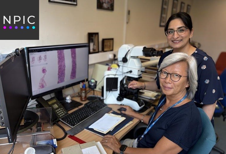
Kirsty Geldeart and Chloe Knowles, lab technician and biomedical scientist at Leeds Teaching Hospitals NHS Trust (LTHT), have put together their #TopLabTips for whole slide imaging in the laboratory.
Firstly, All labs are different and these tips help our team improve workflow and maintaining our equipment.
Number One
Remember to make sure slides are dry before putting them into scanner racks. If the mountant is still wet after staining and coverslipping, then the slides will get stuck in the rack.

Number Two
Make sure tissue sections aren’t too thick. When sectioning on the microtome, the sections are 3 microns is standard for us and the thicker it is the harder it is for the scanner to focus and get a good quality image.
Number Three
Making sure the slides are clean before going into scanner racks. If they are dirty or have a build-up of wax, the scanner may identify it as tissue and try to scan it. Also, debris in the scanner falls off from the slide and can damage the internal elements of scanning equipment.

Number Four
To make it easier to case up the slides after scanning, in Leeds Histo we leave a gap between slides in the rack when the cases start/end to ensure we don’t miss anything.

Number Five
Remembering to check that there is no dust on the lens inside the scanner, we do a light wipe with a Q-Tip to remove any dust.